FREE DOWNLOAD
Windows: https://cdn.toltech.net/sds/downloads/visible-brain-atlas/6.4.8/visible-brain-atlas.exe
Mac: https://cdn.toltech.net/sds/downloads/visible-brain-atlas/6.4.8/visible-brain-atlas.zip

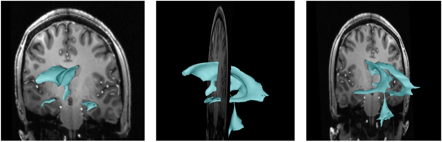
The Visible Brain Atlas is an open source software program created by Touch of Life Technologies in collaboration with Dr. Maureen Stabio, Associate Professor at University of Colorado School of Medicine and several of her amazing MHA students. It includes a high resolution 7 Tesla MRI dataset of the brain (actually, Dr. Stabio’s brain to be exact!) with 3D object overlays of internal structures, ventricular system, cortical parcellation, and vasculature. One of the top things students struggle with the most in neuroanatomy is learning how to read an MRI and visualizing the 3D structures based off of the 2D slices. Now teaching these spatial relationships is as easy as 1-2-3, because students can overlay 3D object such as the ventricles, hippocampus, amygdala, thalamus, hypothalamus and basal ganglia onto the MRI slices. The 3D structures can be toggled on and off, highlighted and labeled while the brain is sliced in any plane. Many out to my current and former students, Emily Mastej, Jake Shearer, and Michael Corigliano from the Modern Human Anatomy Program for the segmentations! Sneak peak on how to use the Visible Brain here.
Does this really help? The efficacy of this teaching approach for training medical students to read MRIs of the brain was evaluated among first year medical students and published in Anatomical Sciences Education. The study showed that this method improves students’ ability to identify structures (particularly complex c-shaped structures) on a 2D MRI slice compared to conventional teaching tools.
 This work is licensed under a Creative Commons Attribution-NonCommercial-ShareAlike 4.0 International License.